
MAIN
----------
Cranial nerve exits
Cranial nerve functions
Cranial nerve reflexes
Head muscles
Bones of the skull
SCALP
Meninges
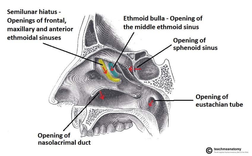
Nasal meati
Nasolacrimal duct
Parasympathetics Video
INFORMATION
-------------------------
Cranial Nerves
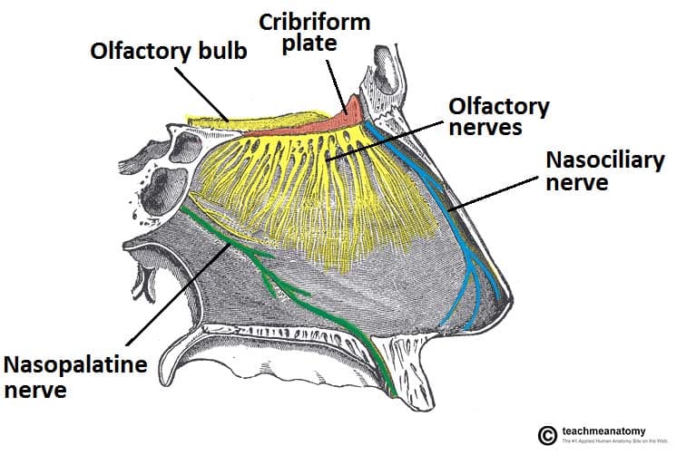
- Olfactory nerve
- Cranial nerve exit -> Cribiform plate of ethmoid bone
- Component ->Special afferent (SA)
- Function -> Sense of smell
- Clinical
- Findings -> Loss of smell (Anosmia)
- Example -> Injury to cribiform plate, congentital absence
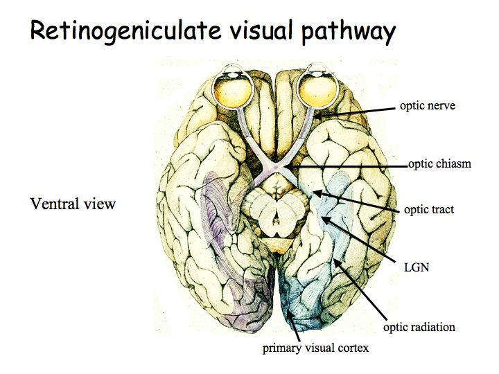
- Optic nerve
- Cranial nerve exit -> Optic canal
- Component ->Special afferent (SA)
- Function -> Eyesight
- Clinical
- Findings -> Blindness, visual field abnormalities, loss of pupillary constriction
- Example -> Direct trauma to the orbit, disruption of optic pathway
- Occulomotor nerve
- Cranial nerve exit -> Superior orbital fissure
- Components ->General Somatic Efferent (GSE), General Visceral Efferent (GVE -parasympathetics)
- Function
- GSE -> Innervation of levator palpebrae superioris, superior, medial, and inferior recti, inferior oblique muscles
- GVE -> Innervation of sphincter pupillae
- Clinical
- Findings -> Dilated pupil, ptosis, loss of normal pupillary reflex, eye moves down inferiorly and laterally (down and out) (H-Test)
- Example -> Pressure from posterior communicating, posterior cerebral, or superior cerebellar artery, pressure from cavernous sinus mass or thrombosis
- Trochlear nerve
- Cranial nerve exit -> Superior orbital fissure
- Component-> General Somatic Efferent (GSE)
- Function -> Innervation of Superior oblique muscle
- Clinical
- Findings -> Inability to look down and in (H-test)
- Example -> lesion along the course of the nerve around the brainstem, orbital fracture
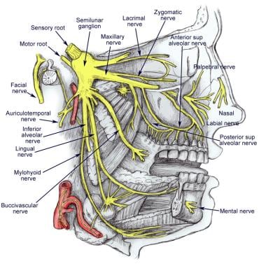
- Trigeminal nerve
- V1 - Opthalmic
- Cranial nerve exit -> Superior orbital fissure
- Component -> General Somatic Afferent (GSA)
- Function -> General sensory from eyes, conjunctiva, orbital contents, nasal cavity, frontal sinus, Ethmoidal air cells, upper eyelid, dorsum of nose, anterior scalp, dura in anterior cranial fossa, Superior part of tentorium cerebelli.
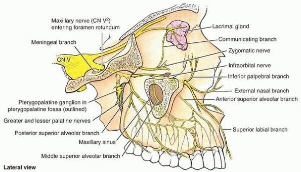
- V2 - Maxillary
- Cranial nerve exit -> Foramen Rotundum
- Component -> General Somatic Afferent (GSA)
- Function -> General sensory from dura in middle cranial fossa, nasopharynx, palate, nasal cavity, upper teeth, maxillary sinus, Skin covering lower eyelids side of nose cheek upper lip,
- V3 - Mandibular
- Cranial nerve exit -> Foramen Ovale
- Component -> General Somatic Afferent (GSA), Special Visceral Efferent (SVE)
- Function
- GSA -> Skin of lower face, anterior part of external ear, part of external acoustic meatus, temporal fossa,anterior 2/3rds of tongue, lower teeth, mucous membranes of cheek, mastoid air cells, mandible
- SVE -> Innervates Temporalis, masseter, medial and lateral pterygoids, tensor tympani, tensor veli palatini, anterior belly of digastric, mylohyoid muscles.
- Clinical
- Findings -> Loss of sensation and pain in region supplied by three divisions of nerve, loss of motor function to muscles of mastication of the side of face of affected V3 nerve
- Example -> Damage to or near specific cranial nerve exit (SOF,FR,FO), lesion in the region of the trigeminal ganglion
- V1 - Opthalmic
- Abducent nerve
- Cranial nerve exit -> Superior orbital fissure
- Component -> General Somatic Efferent (GSE)
- Function -> Innervates lateral rectus muscle
- Clinical
- Findings -> Inability of lateral eye movement (H-test)
- Example -> Brain lesion or cavernous sinus lesion extending into the orbit
- Facial nerve
- Cranial nerve exit(s) -> Internal acoustic meatus -> Stylomastoid foramen
- Components -> General Somatic Afferent (GSA), Special Afferent (SA), General Visceral Efferent (GVE), Special Visceral Efferent (SVE)
- Function
- GSA - Sensory from part of external acoustic meatus and deeper parts of auricle
- SVA - Taste from anterior 2/3rds of tongue
- GVE - Innervates lacrimal, submandibular, and sublingual glands, as well as mucous membranes of nasal cavity, hard and soft palates.
- SVE - Innervation to muscles of facial expression, scalp from 2nd pharyngeal arch, stapedius, posterior belly of digastric, stylohyoid muscles
- Clinical
- Findings -> Paralysis of facial muscles, abnormal taste sensation from anterior 2/3rds of tongue, dry conjunctivae. Paralysis of contralateral facial muscles below the eye.
- Example -> Damage to branches within parotid gland, injury to temporal bone, viral inflammation of nerve, brainstem injury, Bell’s palsy
- Vestibulocochlear nerve
- Cranial nerve exit -> Internal acoustic meatus
- Component -> Special Visceral Afferent
- Functions
- Vestibular division -> Balance
- Cochlear division -> Hearing
- Clinical
- Findings -> Progressive unilateral hearing loss and tinnitus, balance issues.
- Example -> Injury that affects internal acoustic meatus.
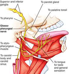
- Glossopharyngeal
- Cranial nerve exit -> Jugular foramen
- Components -> General Visceral Afferent (GVA), Special Visceral Afferent (SVA), General Somatic Afferent (GSA), General Visceral Efferent (GVE), Special Visceral Efferent (SVE)
- Functions
- GVA - Sensory from carotid body and sinus
- GSA - Posterior 1/3rd of tongue, palatine tonsils, oropharynx, and muscosa of middle ear,pharyngotympanic tube, and mastoid air cells
- SVA - Taste from posterior 1/3rd of tongue
- GVE -Innervates parotid salivary gland
- SVE -Innervates stylopharyngeus muscle
- Clinical
- Findings -> Loss of taste and sensation to posterior 1/3rd of tongue, sensation of the soft palate
- Example -> brainstem lesion, penetrating neck injury, damage to jugular foramen
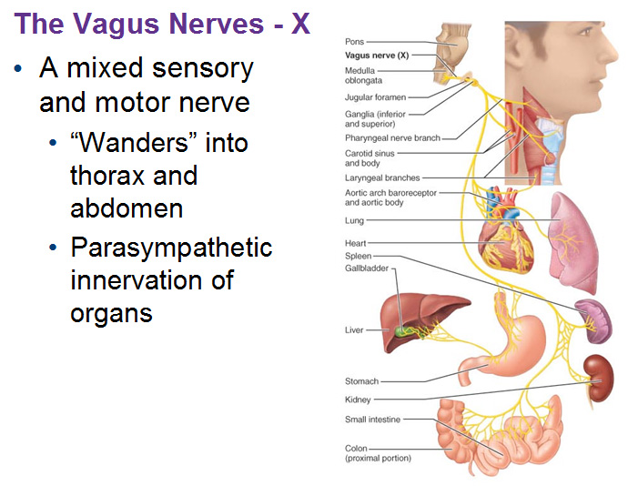
- Vagus nerve
- Cranial nerve exit -> Jugular foramen
- Components ->General Visceral Afferent (GVA), Special Visceral Afferent (SVA), General Somatic Afferent (GSA), General Visceral Efferent (GVE), Special Visceral Efferent (SVE)
- Functions
- GSA -> Sensory from larynx, laryngopharynx, deeper parts of the auricle, part of external acoustic meatus, dura in posterior cranial fossa.
- GVA ->Sensory from aortic body chemoreceptors and baroreceptors, esophagus, bronchi, lungs, heart, abdominal viscera of foregut and midgut.
- SVA -> Taste from epiglottis and pharynx.
- GVE -> Innervates smooth muscle and glands in the pharynx, larynx, thoracic viscera, and abdominal viscera in the foregut and midgut
- SVE -> Innervates palatoglossus, muscles of soft palate except tensor veli palatini, and pharynx except stylopharyngeus, and larynx
- Clinical
- Findings -> Soft palate deviation, deviation of uvula to the UNAFFECTED side, vocal cord paralysis, thoracic and abdominal organ issues
- Example -> Penetrating neck injury, brainstem lesion, damage to jugular foramen
- Accessory Nerve
- Cranial nerve exit -> Jugular foramen
- Component -> General Somatic Efferent (GSE)
- Function -> Innervation of sternocleidomastoid and trapezius muscles
- Clinical
- Findings -> Paralysis of SCN and Trap
- Example -> Penetrating injury to posterior triangle of the neck
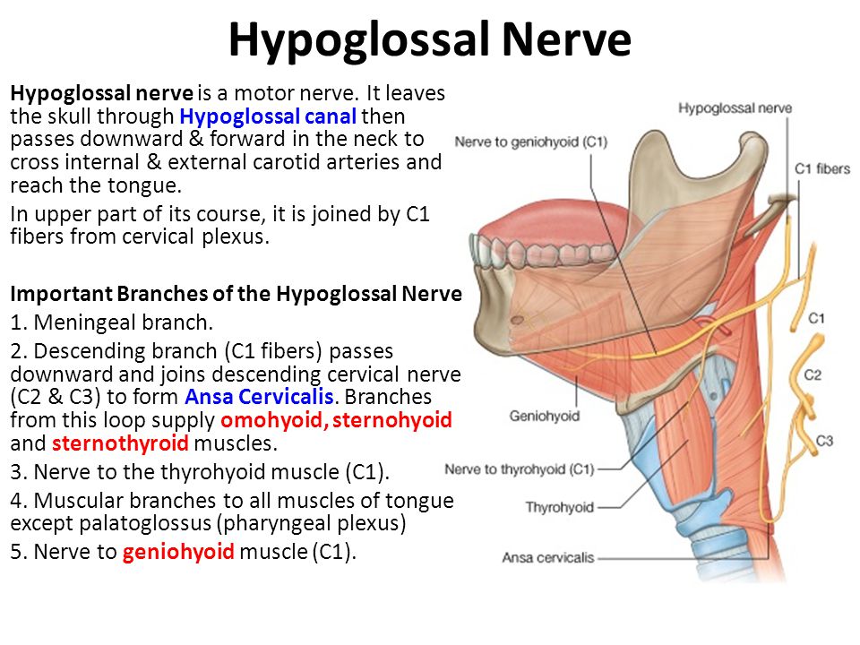
- Hypoglossal Nerve
- Cranial nerve exit -> Hypoglossal canal
- Component -> General Somatic Efferent (GSE)
- Function -> Innervation of hyoglossus, genioglossus, styloglossus, and all intrinsic muscles of the tongue
- Clinical
- Findings -> Atrophy of ipsilateral muscles of the tongue, deviation toward AFFECTED side.
- Example -> Penetrating injury to the neck and skull base pathology
Muscles of the Mastication
|
Muscle |
Origin |
Insertion |
Innervation |
Function |
|
Zygomatic arch and maxillary process |
Lateral surface of ramus of mandible |
Masseteric nerve V3 |
Elevation of mandible |
|
|
Temporal fossa/fascia |
Coronoid process of mandible/ anterior margin of ramus of mandible |
Deep temporal nerves V3 |
Elevation and retraction of mandible |
|
|
Medial surface of lat. plate of pterygoid process Tuberosity of maxilla |
Medial surface of mandible near angle |
Medial pterygoid nerve from V3 |
Elevation and side to side movements of mandible |
|
|
Roof of infratemporal fossa, lateral surface of lat. plate of pterygoid proc. |
Capsule of temporomandibular joint |
Lateral pterygoid nerve from V3 |
Protrusion and side to side movements of mandible |
Muscles of the head
|
Muscle |
Origin |
Insertion |
Innervation |
|
|
Medial palpebral ligament, nasal part of frontal bone, frontal process of maxilla |
Lateral palpebral raphe |
Facial nerve |
Close eyelids |
|
|
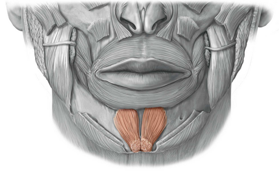
Mandible inferior to incisor teeth |
Skin of chin |
Facial nerve (mental nerve) |
Raises and protrudes lower lip |
|
|
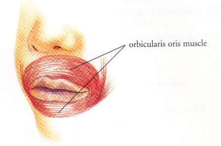
Maxilla and mandible |
Blends with muscle into lips |
Facial nerve |
Closes and protrudes lips |
|
|
Posterior maxilla and mandible, pterygomandibular raphe |
Blends with orbicularis oris |
Facial nerve (buccal nerve) |
Presses cheeks against teeth, compresses distended cheeks |
|
|
Skin of eyebrows |
Scalp aponeurosis |
Facial nerve |
Wrinkles forehead raises eyebrows |
|
|
Lateral superior nuchal line |
Scalp aponeurosis |
Facial nerve |
Draws scalp backwards |
|
|
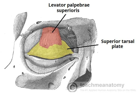
Lesser wing of sphenoid |
Skin of eyelid |
Occulomotor |
Raises eyelid |
|
|
Common tendinous ring |
Superior anterior eyeball |
Occulomotor |
Elevation, adduction, medial rotation of eyeball |
|
|
Common tendinous ring |
inferior anterior eyeball |
Occulomotor |
Depression adduction lateral rotation of eye |
|
|
Common tendinous ring |
medial anterior eyeball |
Occulomotor |
Adduction of eyeball |
|
|
Common tendinous ring |
Lateral anterior eyeball |
Abducent |
Abduction of eyeball |
|
|
Body of sphenoid bone, superior medial to optic canal |
Outer posterior quadrant of eyeball (sup) |
Trochlear |
Depression, abduction, medial rotation of eyeball |
|
|
Medial floor of orbit, maxilla lateral to nasolacrimal groove |
Outer posterior quadrant of eyeball (inf) |
Occulomotor |
Elevation abduction and lateral rotation of eyeball |
---------------------
- External Carotid artery - Some Anatomists Like Freaking Out Poor Med Students.
- Superior Thyroid artery
- Supplies thyroid
- Must be ligated when conducting a thyroidectomy
- Ascending pharyngeal artery
- Supplies the pharynx
- Lingual artery
- Supplies genioglossus
- Facial artery
- Arises in Carotid triangle
- Supplies Facial muscles
- Occipital artery
- Supplies scalp, occipitalis m., SCN m., deep back mm. and neck mm.
- Posterior auricular artery
- Supplies scalp and auricle
- Maxillary artery
- Supplies deep structures to the face
- Middle meningeal arteries
- Clinical -> Epidural hematomas
- Superficial temporal artery
- Supplies temporalis, temple, scalp
- Superior Thyroid artery
- Internal Carotid/Circle of Willis, Blood supply to the brain
- Internal Carotid artery - Enters through the carotid canal to join up with the vertebral artery and form the Circle of Willis
- Vertebral artery - Branch off of the Subclavian artery, runs up vertebral column and enters skull through Foramen magnum
- Venous Drainage
- Superior Sagittal Sinus - receives CSF
- Inferior Sagittal Sinus - Drains into straight sinus
- Straight Sinus - Drains into confluence of sinuses
- Confluence of Sinuses - receives from superior sagittal sinus, straight sinus, and occipital sinuses
- Confluence -> Transverse -> Sigmoid -> Internal Jugular
SCALP and Meninges
-----------------------------
Scalp consists of 5 layers
- Skin - outermost layer, structurally similar to skin throughout the body
- Connective tissue (dense) - Anchors skin to aponeurosis, contains arteries, veins, and nerves supplying the scalp
- Aponeurosis - Consists of occipitofrontalis muscle and the epicranial aponeurosis that connects the two.
- Loose Connective Tissue - Connects aponeurosis to pericranium. Infections tend to spread and localize through the LCT
- Pericranium - Deepest layer of the scalp, periosteum of the outer surface of the calvaria. Can be removed except in areas near the sutures
- Dura mater - Outermost meningeal layer, Periosteal outer layer and Inner meningeal layer. The periosteal layer contains the middle meningeal arteries. Split sagittally by falx cerebelli, axially by tentorium cerebelli
- Arachnoid mater - Middle layer. Avascular, contains arachnoid granules that allow CSF from arachnoid space into the venous system
- Pia mater - Innermost layer, can not be removed from brain.
CLINICAL SIGNIFICANCE
---------------------------------
Hematomas
- Epidural hematoma
- Caused by hemorrhaging of middle meningeal arteries
- Canonical lens shape on CT
- Someone suffering an epidural hematoma may briefly lose consciousness and then regain consciousness.
- Subdural hematoma
- Caused by rupture of bridging veins between dura and arachnoid mater
- Crescent shape on CT
- Someone suffering a subdural hematoma may briefly lose consciousness and then regain consciousness.
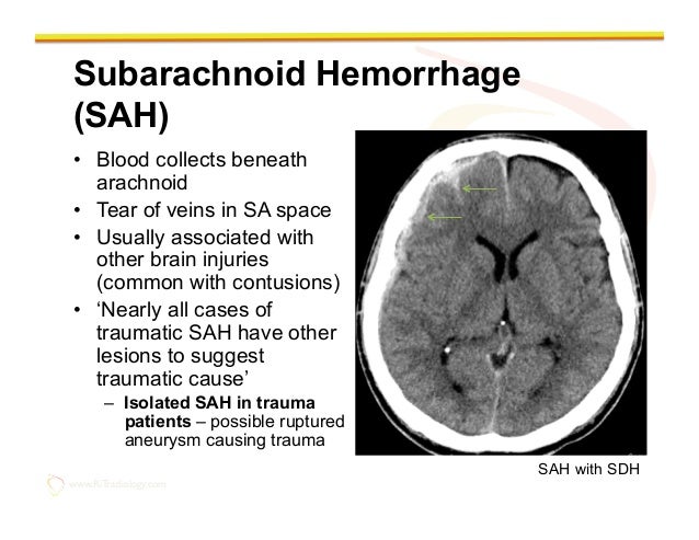
- Subarachnoid hematoma
- Caused by rupture of brain vasculature between arachnoid and pia mater
- “Worst headache I’ve ever had in my life”
- Blown Orbit
- Floor of the orbit fractures usually due to contact with a fast moving object such as a ball or elbow.
- Symptoms include double vision, enopthalmos, loss of sensation in cheek and upper gums
- Cribiform plate fracture
- Trauma to the head may cause a fracture of the cribiform plate of the ethmoid bone.
- 2 common signs of this would be Anosmia (due to olfactory nerve damage), and CSF leakage from the nasal canal.
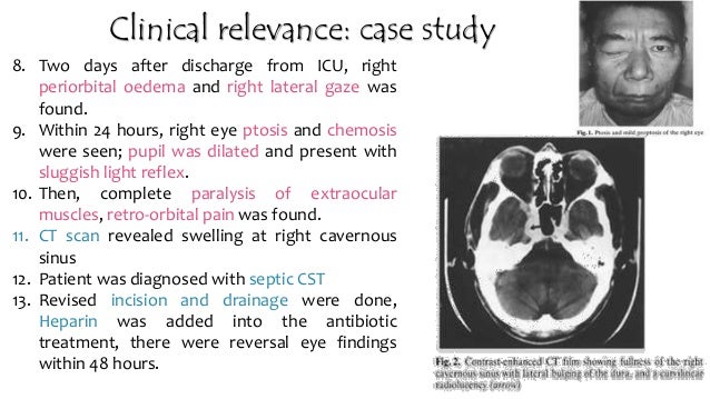
- Caused by coagulation or blood clot in cavernous sinus.
- Common signs are edema, headache, photophobia, and bulging of the eye (proptosis), Ptosis, chemosis (swelling of conjunctiva), cranial nerve palsies (III, IV, V, VI)
- Treated with blood thinners
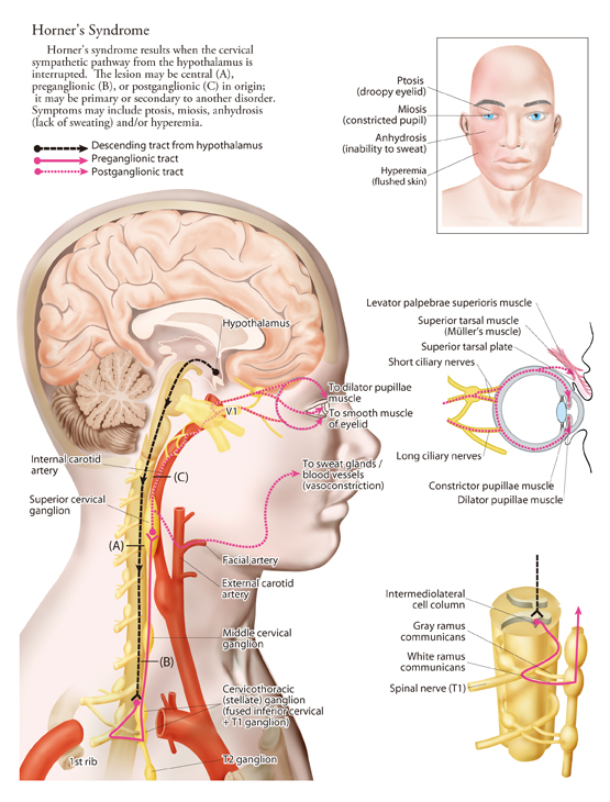
- Symptoms - Ptosis, miosis (dilated pupil), anhydrosis, hyperemia (flushed skin)
- Symptoms - Sweating when hungry, sometimes crying when you eat (really.)
